Echocardiogram: A Simple Guide to Your Heart Ultrasound
Echocardiogram: A Simple Guide to Your Heart Ultrasound
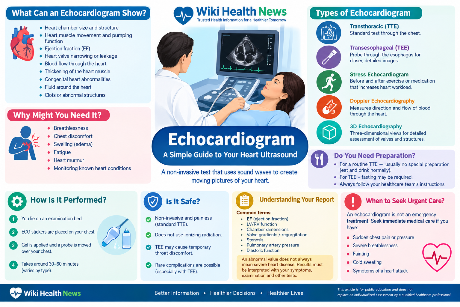
An echocardiogram (echo) is a non-invasive heart ultrasound that uses sound waves to create moving pictures of the heart. It helps doctors assess the heart’s chambers, valves, pumping function, wall motion and blood flow. Unlike an X-ray or CT scan, a standard echocardiogram does not use ionizing radiation. (www.heart.org)
An echocardiogram is commonly used when doctors need to investigate symptoms such as breathlessness, chest discomfort, swelling, fatigue or a heart murmur, and to monitor known heart conditions.
What does an echocardiogram show?
An echo can provide information about:
- Heart chamber size and structure
- Heart muscle movement and pumping function
- Ejection fraction (EF)
- Heart valve narrowing or leakage
- Blood flow through the heart
- Thickening of the heart muscle
- Some congenital heart abnormalities
- Fluid around the heart
- Certain clots or abnormal structures
It is particularly important in evaluating heart failure, valve disease and many other structural heart problems. (www.heart.org)
For patients experiencing possible cardiovascular symptoms, understanding heart attack symptoms and when to seek urgent medical attention is also important.
How is an echocardiogram performed?
The most common examination is a transthoracic echocardiogram (TTE). You usually lie on an examination bed while a sonographer places ECG stickers on your chest and applies ultrasound gel. A handheld probe is moved over different areas of your chest to obtain images of the heart. The examination commonly takes around 30–60 minutes, although the duration varies. (nhs.uk)
The test may be slightly uncomfortable because the probe sometimes needs gentle pressure against the chest, but it is generally painless.
Types of echocardiogram
Different echo techniques may be recommended depending on the clinical question:
Transthoracic echocardiogram (TTE): The standard examination performed through the chest.
Transesophageal echocardiogram (TEE/TOE): A small ultrasound probe is passed into the esophagus to obtain closer, more detailed images of the heart and valves. Sedation and throat numbing are commonly used. (Mayo Clinic)
Stress echocardiogram: Images are obtained before and after exercise or medication that increases the heart’s workload. It can help evaluate how the heart responds to stress.
Doppler echocardiography: Measures the direction and characteristics of blood flow through the heart and valves.
3D echocardiography: Produces three-dimensional views that can be particularly useful for assessing certain valves and structural abnormalities.
Do you need preparation?
For a routine TTE, special preparation is often unnecessary, and many people can eat and drink normally. Preparation can be different for a transesophageal echo, where fasting may be required because the probe passes through the mouth and esophagus. (nhs.uk)
Always follow the instructions provided by your healthcare team.
Is an echocardiogram safe?
A standard echocardiogram is considered a safe, non-invasive test and does not expose you to ionizing radiation. (www.heart.org)
TEE is more involved and may cause temporary throat discomfort. Rare complications can occur, which is why the healthcare team reviews your medical history and explains the procedure beforehand.
Understanding your echo report
An echocardiogram report may contain terms such as EF, LV function, RV function, chamber dimensions, valve gradients, regurgitation, stenosis, pulmonary artery pressure and diastolic function.
An abnormal value does not automatically mean that you have severe heart disease. Results need to be interpreted together with your symptoms, examination, ECG, blood tests and other investigations.
For example, EF is one measure of how effectively the heart pumps blood, but a normal EF does not exclude every type of heart problem. (www.heart.org)
If you have known or suspected heart failure, an echocardiogram may be an important part of assessing cardiac structure and function.
Similarly, people with high blood pressure may eventually develop structural changes in the heart, making appropriate blood-pressure management important.
Echocardiogram vs ECG
An ECG (electrocardiogram) records the heart’s electrical activity, while an echocardiogram produces ultrasound images of the heart’s structure, movement and blood flow. They answer different clinical questions and are often used together.
An echo also does not replace tests designed specifically to assess the coronary arteries. Depending on symptoms and risk factors, doctors may recommend additional testing for coronary artery disease.
When should you seek urgent medical care?
An echocardiogram is an investigation, not an emergency treatment. If you develop sudden chest pressure or pain, severe breathlessness, fainting, cold sweating, or symptoms suggestive of a heart attack, seek emergency medical care rather than waiting for an echocardiogram appointment.
This article is for public education and does not replace an individualized assessment by a qualified healthcare professional.
Author Card
Dr. Sujit Kumar Sahu
Consultant Interventional Cardiologist | Founder & Healthcare Leader | Chief Editor, Wiki Health News
Qualifications: MBBS, MD (Medicine), DNB (Cardiology)
Fellowships/Certifications: FESC, FACC, FSCAI, ICCPR-CRFC
Dr. Sujit Kumar Sahu is a Consultant Interventional Cardiologist and healthcare educator with an interest in cardiovascular prevention, patient education and evidence-based heart health information.
30 Echocardiogram FAQs
1. What is an echocardiogram?
An echocardiogram is an ultrasound examination that creates images of the heart using sound waves.
2. Is an echocardiogram painful?
A standard echocardiogram is generally painless, although pressure from the ultrasound probe may cause mild discomfort.
3. Does an echocardiogram use radiation?
No. Echocardiography uses ultrasound sound waves rather than ionizing radiation. (ASE)
4. How long does an echocardiogram take?
A routine examination commonly takes approximately 30–60 minutes, depending on the type of study.
5. Can I eat before an echocardiogram?
For most standard TTE examinations, normal eating and drinking are usually allowed. TEE generally requires specific fasting instructions. (nhs.uk)
6. What is TTE?
TTE means transthoracic echocardiography, the most common type of echo performed through the chest.
7. What is TEE?
TEE means transesophageal echocardiography. A small ultrasound probe is passed into the esophagus to obtain closer images of the heart. (www.heart.org)
8. What is ejection fraction?
Ejection fraction, or EF, is a measurement used to describe how much blood the ventricle pumps out with each contraction.
9. Can an echo detect heart failure?
Yes. Echocardiography can provide important information about heart structure and pumping function when evaluating heart failure. (www.heart.org)
10. Can an echo detect blocked arteries?
A routine resting echo does not directly show most coronary artery blockages. Additional tests may be needed.
11. Can an echo detect valve disease?
Yes. Echocardiography is a major test for evaluating heart valve structure and function. (www.heart.org)
12. Can an echo detect a heart murmur?
An echo may help identify structural or valve abnormalities responsible for a heart murmur.
13. What is Doppler echocardiography?
Doppler technology evaluates blood flow through the heart and blood vessels.
14. What is a stress echocardiogram?
It evaluates the heart at increased workload, usually after exercise or medication that mimics exercise.
15. Is an echocardiogram safe during pregnancy?
Ultrasound does not use ionizing radiation, but any medical test during pregnancy should be performed when clinically appropriate and interpreted by the healthcare team.
16. Can an echo detect an enlarged heart?
Yes. Echocardiography can measure the size of cardiac chambers and assess structural changes.
17. Can an echo detect a weak heart?
It can identify reduced cardiac pumping function and abnormalities in heart-muscle movement.
18. What does LV function mean?
LV function describes how well the left ventricle, the heart’s main pumping chamber, contracts and functions.
19. What does RV function mean?
RV function describes the performance of the right ventricle, which pumps blood toward the lungs.
20. What does valve regurgitation mean?
Regurgitation means blood leaks backward through a heart valve when it should be closing.
21. What does valve stenosis mean?
Stenosis means a heart valve has become narrowed, restricting normal blood flow.
22. Can an echo diagnose congenital heart disease?
Echocardiography can identify many structural abnormalities present from birth. (www.heart.org)
23. Can an echo show fluid around the heart?
Yes. Echocardiography can identify fluid in the pericardial space.
24. Can an echo detect blood clots?
Certain clots, particularly those in cardiac chambers, may be visible on echocardiography, although detection depends on their location and imaging quality.
25. Do I need an echo every year?
Not necessarily. The frequency depends on your medical condition, symptoms, treatment and your doctor’s assessment.
26. Is an abnormal echocardiogram always dangerous?
No. Some abnormalities are mild or clinically insignificant. The significance depends on the finding and the overall clinical picture.
27. Can a normal echo rule out all heart disease?
No. A normal echo does not exclude every cardiac condition, including some coronary artery and electrical disorders.
28. Who performs an echocardiogram?
A trained sonographer or cardiac imaging professional commonly performs the examination, with interpretation by an appropriately qualified clinician. (nhs.uk)
29. When will I get my echo results?
Timing varies. Some results may be discussed on the same day, while others are reviewed during a later appointment. (nhs.uk)
30. Should I discuss my echo report with my doctor?
Yes. Your clinician can interpret the findings in relation to your symptoms, examination, medical history and other tests.
Trusted References
- American Heart Association — Echocardiogram (Echo)
- American Heart Association — Echocardiogram Patient Fact Sheet
- NHS — Echocardiogram
- Mayo Clinic — Echocardiogram
- American Society of Echocardiography — About Echocardiography
- American Heart Association — Diagnosing Heart Failure
- American Heart Association — Heart Valve Disease Diagnosis and Testing
Suggested SEO details
SEO Title: Echocardiogram: What It Is, Uses, Procedure, Types & Results
Meta Description:
Focus Keyphrase:
Suggested URL Slug: /echocardiogram/








![Lipoprotein(a) [Lp(a)]](https://wikihealthnews.com/wp-content/uploads/2026/08/Understanding_Lipoproteina_Cardiovascular_Risk-scaled-e1787719701457-520x245.png)