ECG and Heart Disease: What an ECG Can Tell You About Your Heart
ECG and Heart Disease: What an ECG Can Tell You About Your Heart
An electrocardiogram (ECG or EKG) is one of the simplest and most commonly used tests for evaluating the heart. It records the heart’s electrical activity and provides information about heart rate, rhythm and electrical conduction. An ECG can help doctors identify several heart conditions, including abnormal heart rhythms, previous or ongoing heart attack, coronary artery disease, cardiomyopathy and some signs of an enlarged or thickened heart.
ECG and Heart Disease: What an ECG Can Tell You About Your Heart
An electrocardiogram (ECG or EKG) is one of the simplest and most commonly used tests for evaluating the heart. It records the heart’s electrical activity and provides information about heart rate, rhythm and electrical conduction. An ECG can help doctors identify several heart conditions, including abnormal heart rhythms, heart attack, coronary artery disease, cardiomyopathy and some signs of an enlarged or thickened heart.
If you experience chest discomfort, pressure, breathlessness, sweating or pain spreading to the arm, jaw or back, it is important to recognize the warning signs of a heart attack and seek urgent medical attention.
What Is an ECG?
An ECG records the electrical signals generated by each heartbeat. During a standard 12-lead ECG, electrodes are attached to the chest and limbs. The test is quick, painless and does not send electricity into the body.
The ECG tracing helps healthcare professionals assess:
- Heart rate
- Heart rhythm
- Electrical conduction
- Cardiac chamber enlargement
- Abnormal electrical patterns
- Changes that may suggest myocardial ischemia or infarction
What Heart Diseases Can an ECG Detect?
1. Heart Attack
An ECG is an important test when a heart attack is suspected. Certain patterns, including ST-segment elevation or depression and T-wave abnormalities, may indicate acute myocardial ischemia or myocardial infarction.
An ECG is not always sufficient to diagnose a heart attack by itself. Doctors generally consider the ECG together with symptoms, serial ECGs, cardiac troponin testing and other clinical findings.
A blood test called high-sensitivity troponin (hs-cTnI/hs-cTnT) can help detect heart muscle injury and is an important part of the evaluation of suspected acute coronary syndrome.
2. Coronary Artery Disease
An ECG may show evidence of myocardial ischemia, previous myocardial infarction or other changes associated with coronary disease. However, a normal resting ECG does not completely rule out coronary artery disease.
People with risk factors such as diabetes, smoking, high cholesterol and high blood pressure may require cardiovascular risk assessment even when their ECG is normal.
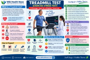
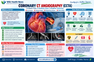
Depending on symptoms and cardiovascular risk, doctors may recommend additional investigations such as an exercise stress test, echocardiography, CT coronary angiography or other cardiac tests.
3. Abnormal Heart Rhythms
ECG is particularly useful for diagnosing arrhythmias such as:
- Atrial fibrillation
- Atrial flutter
- Supraventricular tachycardia
- Ventricular tachycardia
- Bradycardia
- Heart block
- Premature atrial or ventricular beats
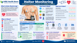
Because a standard ECG records only a short period of heart activity, intermittent symptoms may require longer-term monitoring such as a Holter monitor or event recorder.
4. Left Ventricular Hypertrophy
Long-standing high blood pressure and certain valve or structural heart diseases can cause thickening of the heart muscle. An ECG can provide clues to left ventricular hypertrophy, although an echocardiogram is generally more useful for directly assessing heart structure.
5. Cardiomyopathy
Some cardiomyopathies produce characteristic electrical abnormalities. An ECG can therefore be an important part of the initial assessment. Additional investigations, including echocardiography or cardiac MRI, may be required depending on the suspected condition.
6. Heart Failure
An ECG does not directly measure the pumping strength of the heart, but it can identify rhythm disturbances, previous myocardial infarction, conduction abnormalities and other findings that may accompany or contribute to heart failure.
For more information about symptoms, causes, diagnosis and treatment, see our guide to heart failure.
ECG Changes That May Be Important
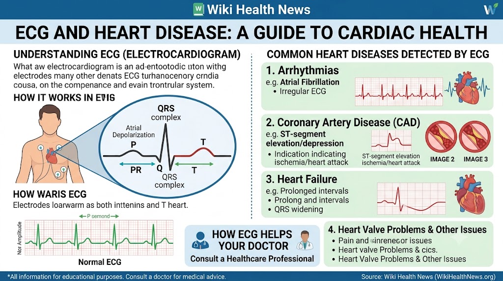
Doctors may look for abnormalities involving different parts of the ECG:
ST segment: Changes may suggest myocardial ischemia or acute myocardial injury.
T waves: T-wave inversion and other abnormalities can occur with ischemia and several other conditions.
Q waves: Certain pathological Q-wave patterns may suggest previous myocardial infarction.
PR interval: Abnormalities can indicate conduction problems.
QRS complex: Changes in width or morphology may suggest bundle branch block, ventricular hypertrophy or other conduction abnormalities.
QT interval: QT prolongation or shortening can be clinically important because it may increase the risk of certain ventricular arrhythmias.
ECG interpretation should always consider the patient’s symptoms, medical history, medications and previous ECGs. An automated ECG interpretation should not be considered a final diagnosis without appropriate clinical review.
Can a Normal ECG Rule Out Heart Disease?
No.
A normal ECG is reassuring but does not exclude every type of heart disease.
For example, some people with coronary artery disease may have a normal resting ECG. Similarly, intermittent arrhythmias may not occur during the few seconds when a standard ECG is recorded.
Depending on the clinical situation, doctors may recommend:
- Repeat or serial ECGs
- High-sensitivity troponin testing
- Echocardiography
- Holter monitoring
- Exercise stress testing
- CT coronary angiography
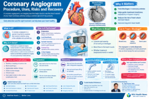
- Coronary angiography
- Cardiac MRI
For selected patients without symptoms, coronary artery calcium assessment may also be considered as part of cardiovascular risk evaluation. A CT calcium score can help estimate the amount of calcified plaque in the coronary arteries, but it is different from an ECG and does not replace evaluation of acute chest pain.
ECG vs Echocardiogram
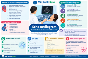
An ECG records electrical activity, while an echocardiogram uses ultrasound to assess the heart’s structure and function.
An ECG is particularly useful for evaluating:
- Heart rhythm
- Electrical conduction
- Heart rate
- Ischemic ECG changes
- Evidence of previous myocardial injury
An echocardiogram provides information about:
- Heart chamber size
- Heart muscle movement
- Ejection fraction
- Heart valves
- Pericardial effusion
- Structural abnormalities
The two tests complement each other rather than replace one another.
When Should You Get an ECG?
A doctor may recommend an ECG if you have:
- Chest pain or pressure
- Palpitations
- Unexplained shortness of breath
- Dizziness or fainting
- Irregular heartbeat
- Suspected heart attack
- Suspected arrhythmia
- Known heart disease
- Certain medication-related cardiac risks
- Cardiovascular risk factors
An ECG may be particularly important when symptoms suggest acute coronary syndrome.
ECG and Chest Pain: Don’t Ignore Warning Signs
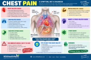
Chest pressure, squeezing or pain that spreads to the arm, shoulder, back, neck or jaw—especially when associated with sweating, nausea or breathlessness—can be a warning sign of a heart attack.
If these symptoms occur, do not wait for a routine ECG appointment or attempt to interpret an ECG yourself. Seek emergency medical care immediately.
An ECG, cardiac troponin testing and other investigations can help doctors determine whether a heart attack or another serious cardiac condition is occurring.
Key Takeaways
- An ECG is a quick, painless and widely available cardiac test.
- It records the heart’s electrical activity.
- ECG is particularly useful for detecting arrhythmias and electrical conduction abnormalities.
- ECG can provide important evidence of acute or previous myocardial infarction.
- A normal ECG does not completely rule out coronary artery disease.
- ECG findings should be interpreted in the context of symptoms and other investigations.
- Depending on the clinical situation, ECG may be combined with troponin testing, echocardiography, stress testing, CT or coronary angiography.
- CT calcium scoring and ECG provide different types of information and should not be considered interchangeable tests.
Medical Disclaimer: This article is intended for general educational purposes and does not replace consultation with a qualified healthcare professional. Anyone with symptoms suggestive of a heart attack should seek emergency medical care immediately.
About the Author
Dr. Sujit Kumar Sahu
Consultant Interventional Cardiologist
Dr. Sujit Kumar Sahu is a Consultant Interventional Cardiologist with expertise in cardiovascular disease, coronary interventions and contemporary cardiac care. His qualifications include MBBS, MD (General Medicine) and DrNB (Cardiology).
He contributes evidence-based cardiology and heart-health education designed to help patients better understand cardiovascular conditions, diagnostic tests and preventive care.
Medical content reviewed by: Dr. Sujit Kumar Sahu
Specialty: Interventional Cardiology
1. What is an ECG?
An ECG is a test that records the electrical activity of the heart.
2. What does an ECG show?
It can show heart rate, rhythm, electrical conduction and several abnormal electrical patterns.
3. Can an ECG detect a heart attack?
Yes. ECG can provide important evidence of an acute heart attack, although doctors usually combine ECG findings with symptoms and cardiac blood tests.
4. Can an ECG detect blocked arteries?
An ECG may show evidence suggesting ischemia or previous heart damage, but a normal ECG cannot reliably exclude blocked coronary arteries.
5. Can a normal ECG mean my heart is healthy?
Not necessarily. Some heart conditions can exist despite a normal resting ECG.
6. Can ECG detect coronary artery disease?
It can provide clues, but additional testing may be required to diagnose or assess coronary artery disease.
7. Can ECG detect atrial fibrillation?
Yes. An ECG can identify atrial fibrillation when the arrhythmia is present during recording.
8. What if palpitations occur but the ECG is normal?
If symptoms are intermittent, a Holter monitor or another ambulatory cardiac monitor may be recommended.
9. Is an ECG painful?
No. A standard ECG is painless.
10. How long does an ECG take?
The actual recording usually takes only a few minutes.
11. What is a 12-lead ECG?
A 12-lead ECG records the heart’s electrical activity from multiple viewpoints using electrodes placed on the limbs and chest.
12. Can ECG detect heart failure?
An ECG can identify abnormalities associated with heart failure, but it does not directly measure the heart’s pumping function.
13. Can ECG detect an enlarged heart?
An ECG may provide clues to cardiac chamber enlargement or ventricular hypertrophy, but imaging such as echocardiography is usually more informative.
14. Can high blood pressure affect an ECG?
Long-standing hypertension can cause changes such as left ventricular hypertrophy that may be reflected on an ECG.
15. What does ST elevation mean?
ST elevation can be associated with acute myocardial injury and, in the appropriate clinical setting, STEMI. It can also occur in other conditions, so interpretation requires clinical assessment.
16. What does ST depression mean?
ST depression can occur with myocardial ischemia but has several possible causes and must be interpreted in context.
17. What does T-wave inversion mean?
T-wave inversion can occur with ischemia and other cardiac or non-cardiac conditions. Its significance depends on the ECG leads and clinical situation.
18. What are Q waves on an ECG?
Certain abnormal Q-wave patterns can indicate previous myocardial infarction or myocardial scar.
19. Can ECG detect heart block?
Yes. Various types of conduction block can produce characteristic ECG findings.
20. Can ECG detect a slow heartbeat?
Yes. An ECG can identify bradycardia and help determine its rhythm and conduction pattern.
21. Can ECG detect a fast heartbeat?
Yes. It can identify and help classify tachycardias.
22. Is ECG safe?
A standard ECG is considered very safe and does not send electrical current into the body.
23. Is ECG enough to diagnose heart disease?
Not always. Depending on symptoms, doctors may need blood tests, echocardiography, stress testing, CT, MRI or angiography.
24. Should I get an ECG for chest pain?
Chest pain should be medically assessed, particularly if it is severe, persistent or associated with breathlessness, sweating, nausea or pain radiating to the arm, jaw or back.
25. Can ECG detect a previous heart attack?
It can show patterns that may suggest a previous myocardial infarction, although interpretation must consider the complete clinical picture.
26. What is the difference between ECG and echocardiography?
ECG assesses electrical activity; echocardiography uses ultrasound to assess heart structure and function.
27. What is the difference between ECG and Holter monitoring?
A standard ECG is a short recording, while a Holter monitor records the heart rhythm continuously for an extended period, often 24 hours or longer.
28. Can an ECG detect all heart problems?
No. Some structural, coronary and intermittent conditions require additional tests.
29. Should an abnormal ECG always be treated?
No. An abnormal ECG is a finding, not necessarily a diagnosis requiring treatment. The cause and clinical context must be assessed.
30. When is an ECG an emergency test?
An ECG is particularly urgent when acute coronary syndrome or a serious arrhythmia is suspected, especially in someone with concerning symptoms.
References
References
- American Heart Association — Electrocardiogram (EKG or ECG)
American Heart Association — Electrocardiogram - American Heart Association — Diagnosing a Heart Attack
American Heart Association — Diagnosing a Heart Attack - American Heart Association — Diagnosing Heart Failure
American Heart Association — Diagnosing Heart Failure - NHS — Electrocardiogram (ECG)
NHS — Electrocardiogram - NHS — Coronary Heart Disease: Diagnosis
NHS — Coronary Heart Disease: Diagnosis - AHA/ACC — Guideline for the Evaluation and Diagnosis of Chest Pain
AHA/ACC Chest Pain Guideline
What is an ECG?
An electrocardiogram (ECG/EKG) records the electrical activity of your heart over a period of time using small sensor pads (electrodes) placed on the skin. It traces the electrical pulses that make the heart beat and pump blood.
What an ECG Can Detect
- Arrhythmias: Irregular heart rhythms, such as Atrial Fibrillation (AFib) or tachycardia.
- Heart Attack (Myocardial Infarction): Evidence of ongoing or past heart muscle damage.
- Conduction Problems: Blockages or delays in the electrical pathways that coordinate heartbeats.
- Hypertrophy: Thickening or enlargement of the heart muscle walls.
Heart Attack Signs on an ECG
- ST-Segment Elevation (STEMI): Indicates a full blockage of a coronary artery requiring immediate emergency intervention.
- ST-Segment Depression / T-Wave Inversion: May indicate partial blockages or reduced blood flow (ischemia) to the heart muscle.
Important: Normal ECG $\neq$ No Heart Disease
A standard ECG only captures a snapshot of your heart’s electrical activity over a few seconds while you are at rest. You can still have severe coronary artery disease or intermittent arrhythmias despite a normal resting ECG.
Additional Cardiac Diagnostic Tests
- Troponin Blood Test: Detects cardiac proteins released into the bloodstream during heart muscle injury.
- Echocardiogram (Echo): An ultrasound scan to view heart valves and pumping function.
- Holter Monitor: A portable ECG device worn for 24–48 hours to record continuous heart rhythms.
- Stress Test: Evaluates how the heart performs under physical exertion on a treadmill or bike.
- CT Coronary Angiography: Advanced imaging to visualize blood vessels and detect plaque buildup.
🚨 Emergency Warning
If you experience severe chest pressure, tightness, or pain accompanied by breathlessness, cold sweating, or pain radiating to your jaw, neck, back, or arm, call emergency services immediately. Do not drive yourself to the hospital.